
Fig. 4.
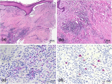
Histopathology of the skin assessed near the necrotic border of digitus 3 of the left hand (Fig. 3) predominantly revealed altered tissue with fibrosis and strong mainly perivascular inflammatory infiltrates including lymphocytes, plasma cells, and neutrophils (a, b). The inflammatory infiltrate was dominated by CD4+ cells (c) when compared to CD8+ lymphocytes (d)