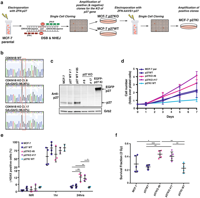
Figure 5.

Generation of MCF-7 luminal breast cancer cells KO and KI for p27. (a) Schematic representation of the experimental workflow used to generate MCF-7 p27KO and KI clones. Briefly, MCF-7 cells were electroporated to incorporate vectors encoding for engineered ZFNs cutting at the CDKN1B gene. Consequent events of DSB and NHEJ were exploited to obtain p27KO clones. Following validation of CDKN1B gene KO, the identified clone was further electroporated with vectors encoding for engineered ZFNs, annealing on the AAVS1 safe harbor gene. The reaction was carried out in the presence of p27WT DNA sequence (fused to EGFP), to induce the generation of p27KI clones through integration. (b) Representative images of the DNA sequence, evaluated by Sanger method, corresponding to CDKN1B gene, showing the WT and the mutated sequences in two p27KO clones (#8 and #17). The two clones both display a single base deletion, confirmed also by NGS. (c) Western Blot analysis of p27 expression of two WT, two p27KO and 1 EGFP p27KI clone. Grb2 was used as loading control. (d) Growth curve analysis of above described clones. Cells were plated in 6-well plates and counted every other day, for 7 days. Data are expressed as fold increase over the number of plated cells (5 × 10⁴) and represent the mean of three independent experiments performed in duplicates, with SD. (e) Graph reports the quantification of the γH2AX positive cells in NIR cell clones at 1 hour and 24 hours after 2 Gy. (f) Graph reports the survival fraction of the different MCF-7 cell clones subjected to 2 Gy and then plated in clonogenic assay. ZFN, zinc fingers nucleases; DSB, double strand breaks; NHEJ, non-homologous end joining; AAVS1, Adeno-Associated Virus Integration Site 1; EGFP, enhanced green fluorescent protein. *Indicates a p < 0.05; **p < 0.01; ***p < 0.001.
