Figure 1.

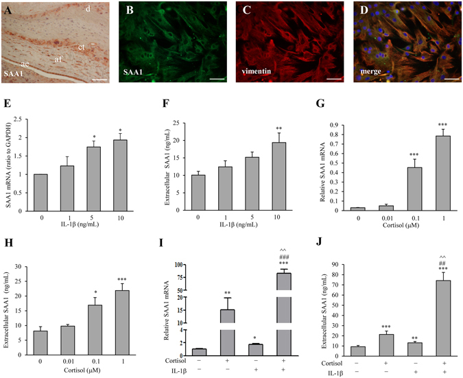
Expression of SAA1 in human fetal membranes and induction of SAA1 production by cortisol and IL-1β. (A) Immunohistochemical staining of SAA1 in human fetal membranes. ae: amnion epithelial cells; af: amnion fibroblasts; ct: chorionic trophoblasts; d: decidua. Scale bar, 200 µm. (B–D) Immunofluorescence staining of SAA1 (B, green) and vimentin (C, red), a marker of mesenchymal cells, in human amnion fibroblasts. Yellow color after merging B and C represents merged staining for SAA1 and vimentin (D). Nuclei were counterstained blue with DAPI. Scale bar, 100 µm. (E–H) The effect of IL-1β (1, 5 and 10 ng/mL, 24 hours) and cortisol (0.01, 0.1 and 1 µM, 24 hours) on SAA1 mRNA (E and G) and secreted SAA1 protein (F and H) in human amnion fibroblasts. n = 4. (I,J) The synergistical effect of IL-1β (10 ng/mL) and cortisol (1 µM) on SAA1 mRNA (G) and secreted SAA1 protein (J) in human amnion fibroblasts. n = 4. Data are the means ± SEM. Statistical analysis was performed with one-way ANOVA test (E–J). *P < 0.05, **P < 0.01, ***P < 0.001 vs. control (0), ##P < 0.01, ###P < 0.001 vs. IL-1β. ^^P < 0.01 vs. cortisol.
