Figure 4.

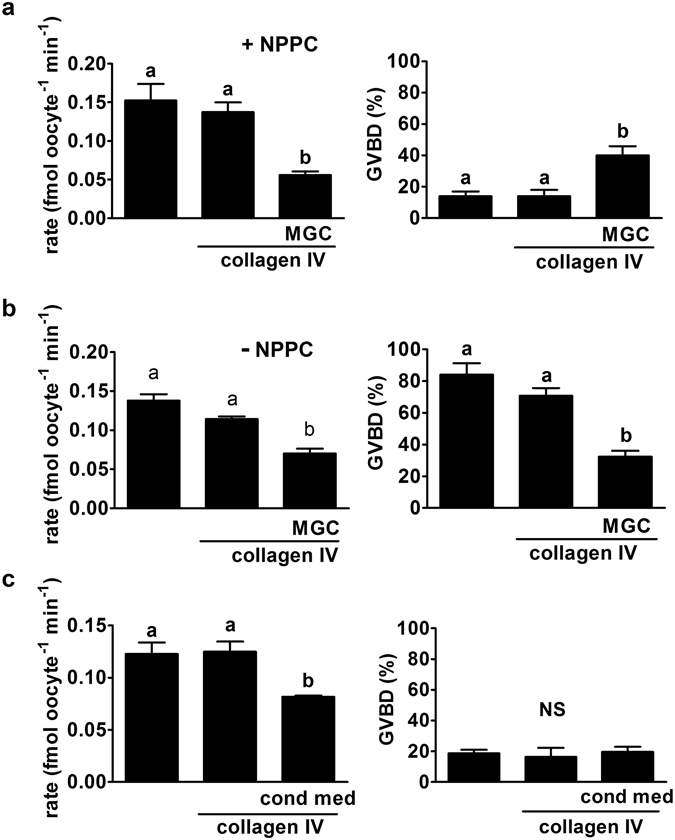
Effect of co-culture of COCs on MGC monolayers. (a) COCs were cultured for 4 h in plastic wells, on collagen IV alone, or on a MGC monolayer on collagen IV as indicated at bottom. Oocytes were then isolated, GV status determined (right) and GLYT1 activity measured (left). GLYT1 was similarly activated within COCs on plastic alone or on collagen IV, but the development of activity was significantly suppressed in the presence of MGC (overall: P = 0.007; a vs b: P < 0.05). GVBD was inhibited in all three groups, since NPPC was present during COC culture, although there was a small but significant increase in the presence of MGC (overall P = 0.009; a vs. b: P < 0.05). Each bar represents the mean ± s.e.m. of three independent repeats. Throughout (a–c), data were analysed by ANOVA with Tukey test. (b) The same groups were again assessed, except that NPPC was absent. MGC again suppressed GLYT1 activity (left; overall, P < 0.0001; a vs. b: P < 0.001) and were also sufficient to inhibit GVBD (right: overall, P < 0.0001; a vs. b: P < 0.001) even in the absence of exogenous NPPC. Each bar represents the mean ± s.e.m. of five independent repeats. (c) The effect of conditioned media from each of the treatment groups in (A) with NPPC present during COC culture was assessed. Conditioned medium from MGC, but not plastic or collagen IV alone, significantly suppressed GLYT1 activation (left; (overall P = 0.018; a vs. b: P < 0.05). GVBD was equally prevented in all three groups (right; P = 0.84; not significant, NS). Each bar represents the mean ± s.e.m. of three independent repeats.
