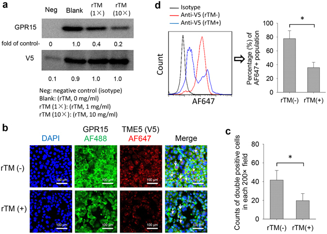
Figure 1.

TME5 interacts with GPR15. (a) Immunoprecipitation. Membrane proteins isolated from HUVECs were incubated with V5-tagged TME5 (100 μg/ml) in the presence of gradient doses of rTM (0 mg/ml, 1 mg/ml, 10 mg/ml), followed by immunoprecipitation with anti-V5 antibody. Mouse derived isotype antibody was used as negative control. The precipitated proteins were subjected to Western blot analysis to detect the indicated proteins. Figure represents one of the three independent experiments. Relative intensities of bands were analyzed with ImageJ software. (b–d) EA.hy926 cells were pre-incubated with either rTM or control diluent followed by incubation with V5-tagged TME5 overnight. (b) Immunocytochemistry. Cells were fixed on slide glasses by Autosmear. The fixed cells were stained with anti-GPR15 and V5 antibodies followed by staining with Alexa Fluor 488- and Alexa Fluor 647-conjugated second antibodies. The nuclei were stained with 4′,6-Diamidino-2-phenylindole dihydrochloride (blue). (c) Alexa Fluor 488- and Alexa Fluor 647-double positive cells were indicated by white arrows and were counted on 6 random areas under a microscope. (d) Flow cytometric analysis. Cells were incubated with anti-GPR15 and V5 antibodies followed by staining with Alexa Fluor 488- and Alexa Fluor 647-conjugated second antibodies. Cell were analyzed on a flow cytometer. Figure shows percentage of Alexa Fluor 647-positive population gated on Alexa Fluor 488-positive cells (n = 6). rTM, Recombinant human soluble thrombomodulin; TME5, the fifth EGF-like region of thrombomodulin.
