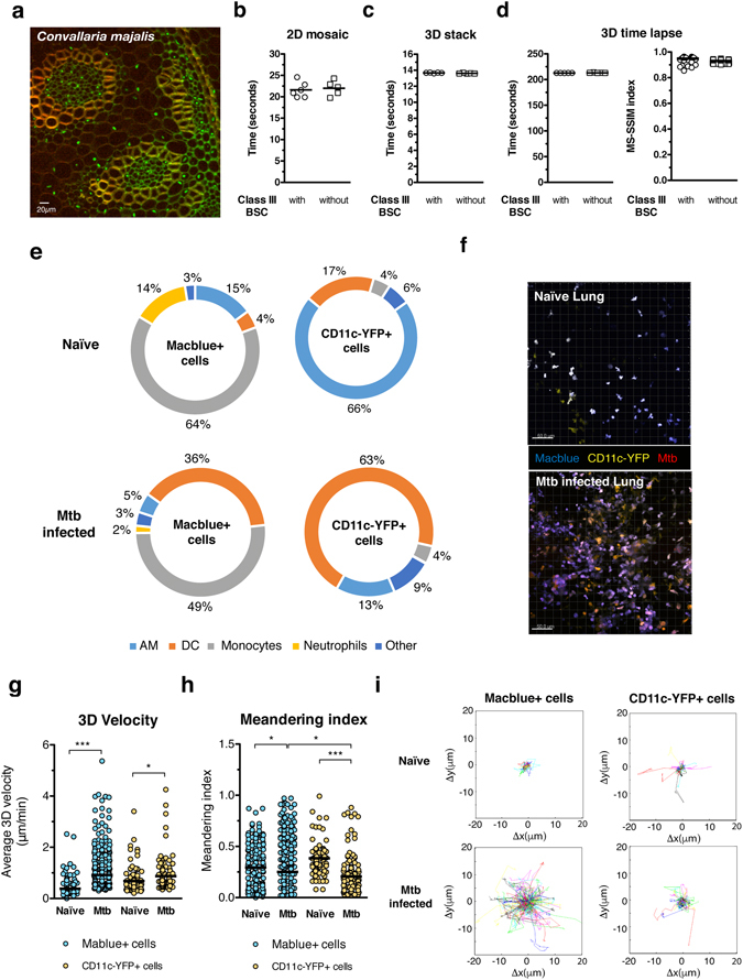
Figure 3.

BSL3 setup for multiphoton microscopy: performance and applications. (a) A Convallaria majalis rhizome slide was used to evaluate the performance of the multiphoton microscope with and without the class III BSC installed. For this images with a size of 505*505 pixels were acquired with 3% of Ti-Saphire laser power tuned to 800 nm and with a pixel dwell time of 2,78 μs. These images were acquired either in 9 different x, y axis positions using the mosaic function (b), 13 different z axis positions with a step of 2 μm using the 3D stack function (c) and 13 z axis positions repeated overtime 15 times (d). Performance of x, y and z axis movement of the microscope was evaluated by recording the time necessary for completion of the different tasks. To quantify image stability overtime we calculated the Structural SIMilarity (SSIM) index between the first 3D stack and each of the subsequent ones within the same time-lapse sequence. Acquisition speed and stability values were calculated from 5 different acquisitions. (e) Flow cytometry analysis of naïve and M. tuberculosis (Mtb) (H37Rv) mouse lung according to the expression of Macblue and CD11c-YFP transgene. (f) Maximum intensity projection of a 3D stack from a naïve and Mtb infected lung vibratome slice. 3D velocity (g), meandering index (h) and Cell Tracks (i) of Macblue+ and CD11c-YFP+ lung cells in vibratome slices. Cell motility parameters were calculated in movies from 3 different areas of lung. Un paired t test was used to evaluate significant differences. *p < 0,05, **p < 0,01, ***p < 0,001.
