
Figure 1.
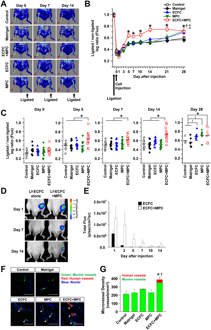
Beneficial effect of human ECFC + MPC on blood flow recovery in the murine ischemic hind limb muscles. Hind limb ischemia was induced by the ligation and cutting of femoral artery and vein at day -1. ECFC, MPC, or ECFC + MPC suspended in Matrigel were injected on day 0. Blood flow was measured by the Laser Doppler imager. Retention of luciferase-labeled ECFC was measured by bioluminescence using the Xenogen IVIS in vivo imaging system. At day 14, perfused human and rat vessels were identified by tail-vein injection of a mixture of rhodamine (red)-conjugated UEA I and FITC (green)-conjugated GS-IB4. (A) Representative laser Doppler images of ischemic hind limbs with/without cell injection over 14 days. (B) Graph of blood flow presented as the ligated/non-ligated leg ratio (n = 11–15; means ± SEM.). *Significant difference (P ≤ 0.05) between groups. (C) Dot graph expressed as single values for each mouse (n = 6–15; means shown by horizontal bars). *Significant difference (P ≤ 0.05) between groups. (D) Representative bioluminescence images of mice after injection of luciferase-labeled ECFC alone or luciferase-labeled ECFC + MPC. (E) Graph of bioluminescence signals detected in ischemic hind limbs (n = 3–4; means ± SEM.). (F) Representative confocal images of lectin-labeled vessels in the ischemic hind limb muscle at day 14 (Scale bars represent 50 μm). White arrowheads point to lectin-labeled (i.e. perfused) vessels. (G) Graph of total, human plus murine microvessels, in the ischemic hind limb muscles with/without cell injections (n = 3; means ± SEM.). *Significant difference (P ≤ 0.05) between groups for total microvessel density. †Significant difference (P ≤ 0.05) between groups for human microvessel density.