


Figure 7.
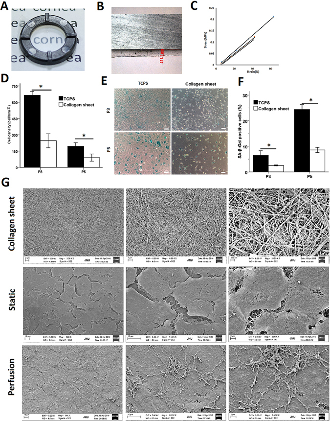
Characterization of the flat collagen sheet and the perfusion system for corneal tissue engineering. (A) Photograph of a flat collagen sheet. (B) Representative section of the collagen sheet. (C) Tensile stress-strain curve for each sample. (D) Cell density was calculated based on the total number of cells in each square millimetre. (E) Cells at different passages on TCPS and on collagen sheets were stained to detect SA-β-Gal activity. Scale bar: 100 μm. (F) The senescence level of the CECs was evaluated based on the ratio of SA-β-Gal-positive cells to total cells. (G) SEM images of the collagen sheet prepared under vitrification conditions and representative images of CECs on collagen sheets in static and perfusion culture. Differences with *P < 0.05 were considered statistically significant.