


Figure 8.
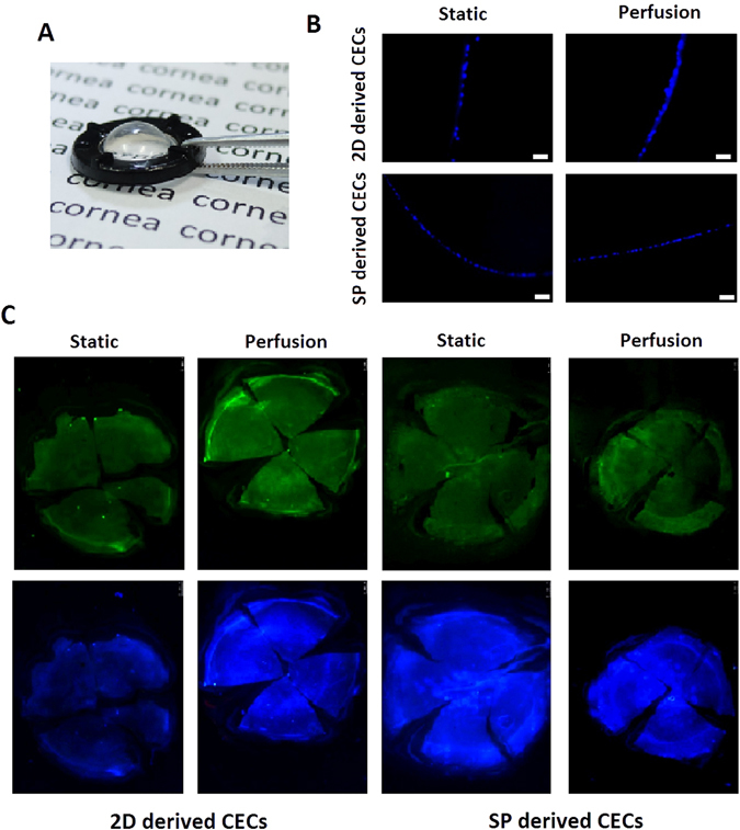
Tissue-engineered CECs on curved collagen sheets in the perfusion system under controlled pressure. (A) Photograph of a curved collagen sheet. (B) Immunohistochemical analysis of cryosectioned spherically curved collagen sheet by DAPI. Scale bar: 50 µm. (C) Representative fluorescence microscopy images of CEC monolayers stained for F-actin and with DAPI on spherically curved collagen sheets in the static and perfusion systems.