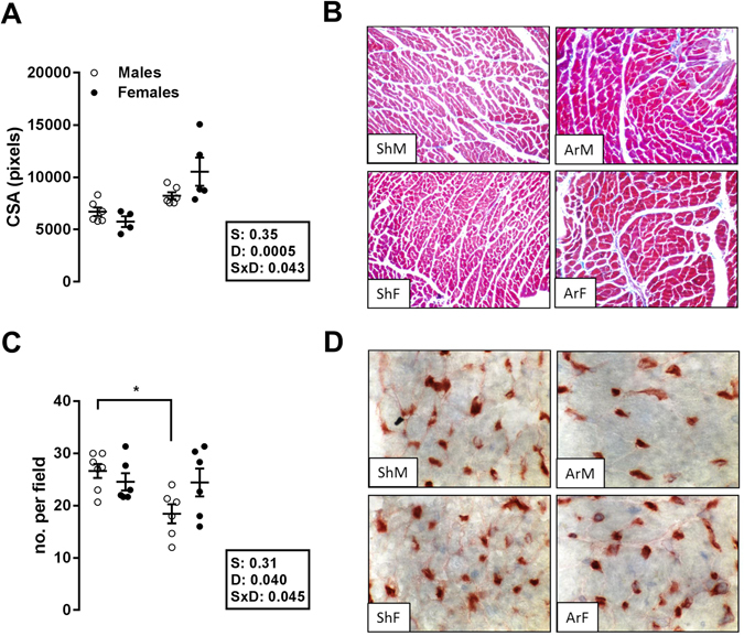
Figure 2.

Impact of LV dilatation and hypertrophy caused by severe aortic regurgitation on cardiomyocyte cross-sectional area (Top panels; A and B) and on myocardial capillary density (bottom panels (C and D). Left panels (A and C): The results are expressed as the mean ± SEM (n = 6–7/gr.). Probability values are from a 2-way ANOVA and Holm-Sedak post-testing. *p < 0.05 or **p < 0.01 between indicated groups. Upper right panels: Representative Masson’s trichrome staining of mid-chamber short-axis LV sections (magnification x200); bottom right panels: LV sections stained with isolectin B4 coupled with horseradish peroxidase (magnification x400).
