Figure 1.

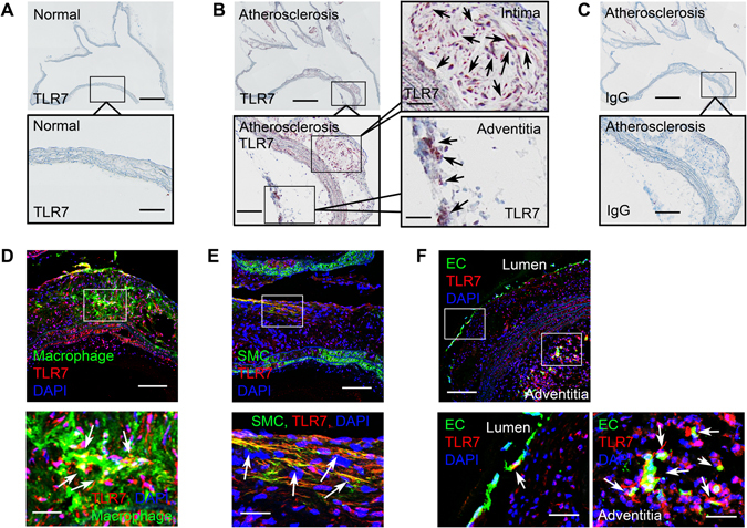
TLR7 expression in mouse atherosclerotic lesions. (A) Rabbit-anti-mouse TLR7 monoclonal antibody immunostaining of a normal mouse aortic arch. Scale: 1 mm, inset scale: 200 µm. (B) Rabbit-anti-mouse TLR7 monoclonal antibody immunostaining of an aortic arch from an Apoe −/− mouse with atherosclerosis showed TLR7 expression in intima and adventitia. Scale: 1 mm, inset scales: 200 and 68 µm. Arrows indicate TLR7-positive cells. (C) Rabbit IgG isotype immunostaining of a parallel aortic arch from an Apoe −/− mouse with atherosclerosis. Scale: 1 mm, inset scale: 200 µm. (D–F) Immunofluorescent double staining of mouse aortic arch atherosclerotic lesions with antibodies against TLR7 and cell type markers (Mac-2, α-actin, and CD31) for macrophage, SMC, and EC from both the lumen and adventitia microvessels. Scale: 100 µm, inset scale: 25 µm. Arrows in the insets indicate TLR7-positive macrophages (D), SMCs (E), and ECs in lumen and adventitia (F).
