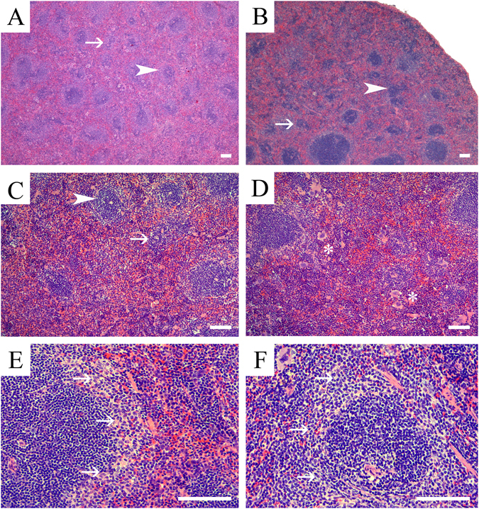
Figure 3.

Representative photomicrographs of splenic microanatomical structures in EE and BPA-exposed adult male and female CD-1 mice. Shown are representative H&E stained sections of splenic tissue from (A) a male mouse exposed to BPA (0.03 ppm), and (B) a female mouse exposed to 3 ppm BPA. These are illustrative of the representative phenotypes in males and females that was characterized by decreased PALS (arrowhead) numbers, size, and lymphocyte cellularity (arrow). These effects were typically more pronounced and diffuse in male versus females. (C) High magnification image of a male spleen representative of the 0.03 ppm BPA exposure group and demonstrating decreased PALS (arrowhead) size, numbers and lymphocyte cellularity (arrow), along with decreased primary lymphoid follicles cellularity (arrow). (D) High magnification image of a representative female spleen from the 3 ppm BPA exposure group characterized by decreased PALS numbers and cellularity with decreased lymphoid follicle cellularity. In addition to the white pulp changes, BPA-exposed females also had increased hematopoietic precursor populations (EMH) within the red pulp, which were predominantly of erythroid lineage (asterisks). Shown is a representative image of a section from a 0.0001 ppm EE-exposed male (E) and female from the 0.03 BPA group (F) showing prominence of marginal zones, a phenotype that was observed in primarily males from EE exposure groups and female from both BPA- and EE-groups. Scale bars indicate 100 µm.
