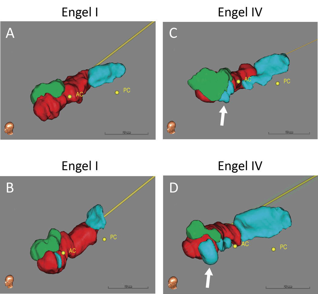
Figure 1. Postoperative ablation volumes.
Volumetric renderings derived from manual tracings of the postoperative ablation zone and remaining non-ablated hippocampal and amygdala tissue, shown for representative Engel I (A and B) and Engel IV (C and D) patients. The white arrows highlight remaining tissue of the mesial hippocampal head in the Engel IV patients. Ablation – red, Amygdala – green, Hippocampaus – blue. AC – anterior commissure, PC – posterior commissure.

