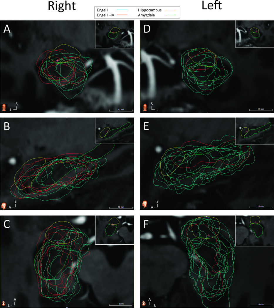
Figure 3. Co-registered ablation cavities and Engel class outcome.
(A) Coronal slice through the right hippocampal head showing greater sparing of medial tissues by the ablations of Engel II–IV patients (red) compared to Engel I patients (blue). Insert in upper right corner demonstrates anatomy, with hippocampus (green) and amygdala (yellow) traced from reference atlas and no ablation cavities shown. (B) Sagittal and (C) axial cuts through right hippocampus and amygdala. The pink crosshairs indicated the intersection of the three imaging planes. (D) Coronal, (E) sagittal and (F) axial slices through left hippocampus with same conventions as (A–C). A – anterior, L – lateral, S – superior.

