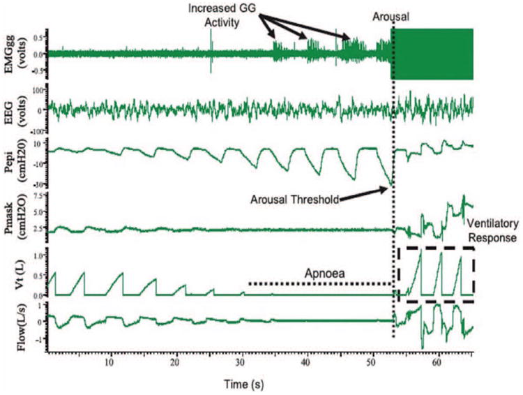
Figure 3.

PSG tracings of an obstructive sleep apnea event in a patient with severe OSA. There was increased EMG activity of the genioglossus muscle during the apneic event, although it was not significant enough to restore flow without arousal. The arousal threshold is characterized using Pepi, which is the epiglottic pressure immediately preceding arousal and there is a large ventilatory response following arousal. Reprinted with permission from Campana et al; Indian J Med Res. 2010;131:176–187. EEG indicates electroencephalogram (C3-A2); EMGgg, electromyogram of the genioglossus muscle (intramuscular); Pepi, pressure at the level of the epiglottis; Pmask, pressure measured via nasal mask; Vt, tidal volume.
