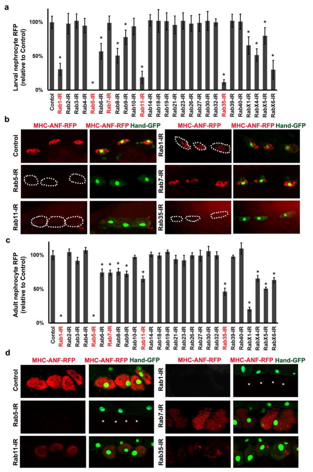
Fig. 3. Nephrocyte uptake of hemolymph protein.
a Second instar larvae nephrocytes in which the indicated Rab gene was silenced were quantitatively assessed for uptake of ANF-RFP fusion protein from the hemolymph. Red fluorescent protein (RFP) fused to rat atrium natriuretic factor (ANF) is expressed in and secreted by muscle cells from a transgene driven by the MHC enhancer. ANF-RFP fluorescence in nephrocytes expressed as percent of WT control. b Fluorescence microscopy showing ANF-RFP uptake (red) by nephrocytes (GFP nuclear expression) in which the indicated Rab gene expression was silenced. Dotted lines indicate outlines of nephrocytes. c Adult flies in which the indicated Rab gene was silenced were quantitatively assessed for uptake of ANF-RFP fusion protein from the hemolymph. ANF-RFP in nephrocytes expressed as percent of WT control. d Fluorescence microscopy showing ANF-RFP (red) uptake by nephrocytes (GFP nuclear expression) in which the indicated Rab gene expression was silenced. Asterisks indicate missing nephrocytes as a result of Rab1 and Rab5 silencing. For quantification, 20 nephrocytes were analyzed from each of 3 larvae or flies per genotype. The results are presented as mean±s.d. Results were analyzed by Student’s t-test. Statistical significance was defined as P<0.05.

