
Figure 1.
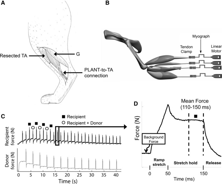
(A) Cartoon of completed PLANT‐to‐TA tendon transfer (medial view) and (B) schematic of experimental set‐up depicting hindlimb muscles detached from their distal insertions and attached in series with myographs and linear motors. During the experiment, two muscles from each limb were connected to separate linear motors and the bone segments were rigidly fixed. (C) Force profiles in response to the ramp stretch‐hold‐release during a crossed extension reflex. In this example, the recipient force is lower when the donor muscle is stretched at the same time consistent with intermuscular force feedback. (D) Example of a single force profile illustrating the mean force time point used for all analyses. TA, tibialis anterior; G, gastrocnemius; PLANT, plantaris