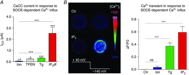
Figure 4. Differential response of Ca2+‐activated Cl− channels to modes of store depletion in the oocyte.

A, amplitudes of current through CaCC following store depletion with different agents: ionomycin, TPEN, thapsigargin (Tg), non‐hydrolyzable IP3 (IP3df). B, left, intracellular Ca2+ transient monitored by confocal microscopy in a voltage‐clamped oocyte using Oregon green BAPTA‐1 under control conditions (Ctr) or following IP3 injection. Ca2+ influx through SOCE was stimulated by hyperpolarizing the cell to −140 mV. Right, summary of the intracellular Ca2+ rise induced by the hyperpolarizing pulse to −140 mV when intracellular stores were depleted with ionomycin (Ion), thapsigargin (Tg) or IP3. Adapted from Courjaret and Machaca (2014).
