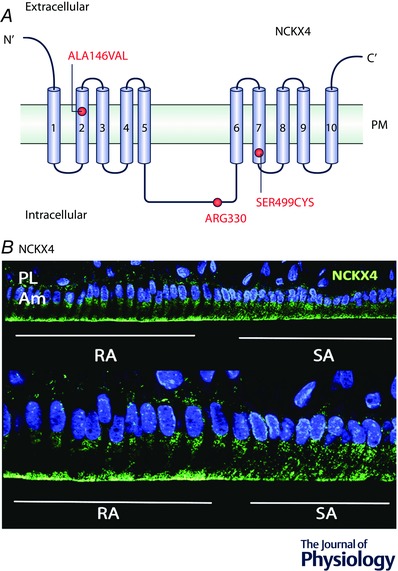
Figure 4. NCKX4 mutations and localization in ameloblasts.

A, schematic representation of NCKX4 protein structure showing the 10 transmembrane domains and corresponding loops. The red dots indicate the mutations at each domain that have been associated with enamel deficiencies. PM = plasma membrane. B, NCKX4 localization in rat ameloblasts. Immunofluorescence microscopy analysis of NCKX4 localization in RA and SA ameloblasts. In RA cells, NCKX4 is largely localized at the apical (distal) pole of the cell. In SA cells, NCKX4 distribution becomes more diffused suggesting a principal role for this protein in Ca2+ extrusion during the RA stage. DAPI is shown in blue. RA = ruffled‐ameloblasts, SA = smooth‐ameloblasts, Am = ameloblasts, PL = papillary layer.
