-
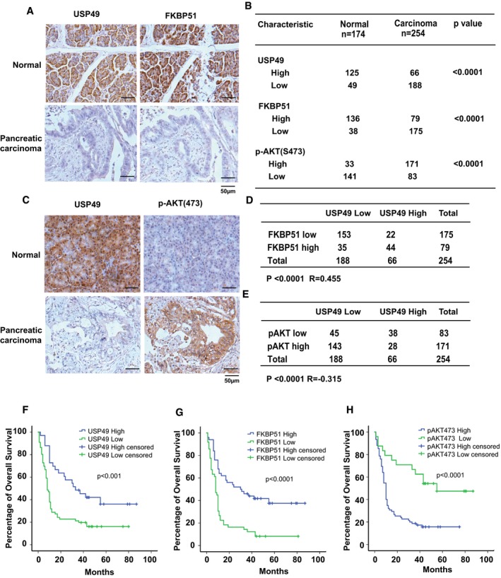
A
Representative images of immunohistochemical staining of USP49 and FKBP51 in normal and pancreatic carcinoma.
-
B
Quantification of USP49, FKBP51, and p‐AKT(S473) protein levels in normal and pancreatic carcinoma. Statistical analyses were performed with the χ2 test.
-
C
Representative images of immunohistochemical staining of USP49 and p‐AKT(S473) in normal and pancreatic carcinoma.
-
D
Correlation study of USP49 and FKBP51 in pancreatic carcinoma. Statistical analyses were performed with the χ2 test. R, Pearson correlation coefficient.
-
E
Correlation study of USP49 and pAKT‐Ser473 in pancreatic carcinoma. Statistical analyses were performed with the χ2 test. R, Pearson correlation coefficient.
-
F–H
Survival analysis of PDAC patients by Kaplan–Meier plots and log‐rank tests. Patients were categorized into high and low expression of USP49, FKBP51, and pAKT‐Ser473 based on IHC staining scores. Censored groups entail non‐cancer deaths or those lost to follow‐up at last recorded follow‐up.