Figure 1.

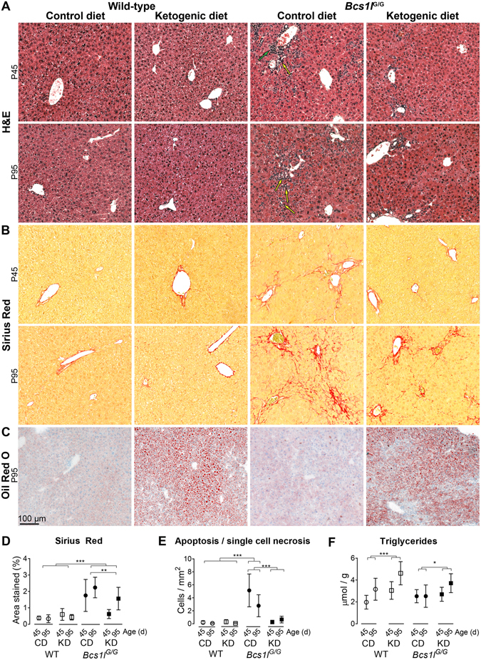
Ketogenic diet modulates hepatopathy progression in Bcs1l G/G mice. (A) Representative H&E stained liver sections showing ductular reactions (yellow arrows) and portal inflammation (green arrow) in the Bcs1l G/G mice on control diet (CD). The ketogenic diet (KD) normalized the expansion of portal areas at postnatal day 45 (P45). A partial amelioration was also observed at postnatal day 95 (P95). For higher-magnification images and their description see Supplementary Fig. 3. (B) Sirius Red staining of liver collagen showing incipient fibrosis at P45 and marked periportal and sinusoidal fibrosis at P95 in the Bcs1l G/G mice on CD. (C) Oil Red O staining of liver cryosections to show neutral lipid accumulation. (D) Quantification of fibrosis from Sirius Red-stained liver sections showing delayed fibrosis in the Bcs1l G/G mice on KD. n = 5–8/group for Bcs1l G/G mice (both time points) and 3–4/group for WT mice at P45 and 8–9 /group at P95, respectively. **p < 0.01, ***p < 0.001 (two-way ANOVA with age group as a factor to be controlled) (E) Count of apoptotic and necrotic cells in H&E-stained liver sections. ***p < 0.001 (Mann-Whitney U-test) (F) Liver triglyceride assay. ***p = 0.0001 for the diet effect, * p = 0.44 for diet*age interaction (two-way ANOVA). n = 7–13/group. The error bars stand for standard deviation. Abbreviations: WT, wild-type mice; Bcs1l G/G, mice homozygous for the Bcs1l c.232A>G knock-in mutation.
