
Figure 2.
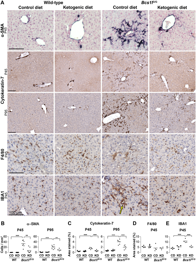
Ketogenic diet modulates activation of hepatic progenitor cells, stellate cells and liver macrophages in Bcs1l G/G mice. (A) Immunostainings of liver sections for hepatic progenitor cell and bile duct marker cytokeratin-7; activated stellate cell marker α-smooth muscle actin (α-SMA) and macrophage markers F4/80 and IBA1. (A,B) In wild-type livers α-SMA staining (dark blue) is seen only in the smooth muscle layer of arterioles but in the Bcs1l G/G mice on CD staining appears in numerous activated spindle-like stellate cells. In the Bcs1l G/G mice on KD the stellate cell α-SMA signal is diminished. (A, C) In wild-type livers cytokeratin-7 staining (dark brown) is only seen in bile ducts whereas in Bcs1l G/G livers the staining is present in numerous cells in the liver parenchyma. The abnormal staining is almost completely absent in the Bcs1l G/G mice on KD at P45. (A,D,E) F4/80 and IBA1 stainings (dark brown) show normal Kupffer cells in wild-type livers. IBA1 staining is increased in Bcs1l G/G livers but normalized by KD. Characteristic IBA1-positive macrophages surrounding a hepatocyte (crown-like structure, arrow) in Bcs1l G/G mice. All scale bars present 100 μm. **p < 0.01; ***p < 0.001.