
Figure 2.
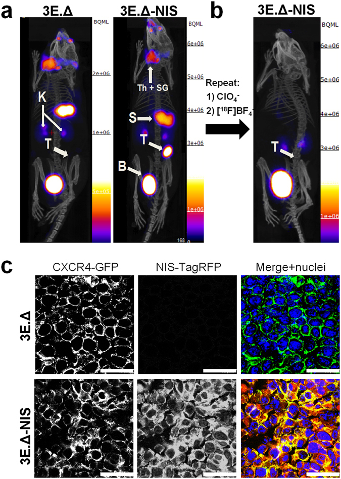
Primary xenograft tumors established from NIS-expressing breast cancer cells can specifically be imaged with [18F]BF4 −-PET. (a) Tumors were established from 3E.Δ cells stably expressing constitutively active truncated CXCR4-GFP but not NIS-TagRFP (left) or from 3E.Δ-NIS cells stably expressing both truncated CXCR4-GFP and NIS-TagRFP (right). Tumors were imaged by [18F]BF4 −-PET when sizes of ~55 mm3 were reached (see Supplementary Fig. S2A). As expected, no tumor signals were seen by PET/CT in the case of 3E.Δ tumors. For 3E.Δ-NIS tumors, PET/CT signals were very pronounced. A representative example is shown. (b) NIS specificity was tested in the same animals 48 h later by pre-treatment with perchlorate before repeat [18F]BF4 −-PET imaging. All [18F]BF4 −-PET signals except in the organs of the [18F]BF4 − excretion pathway, i.e. kidneys and bladder, vanished in perchlorate-treated animals (as compared to the earlier image of the same animal as in (a)), thereby proving successful NIS blockade and thus NIS specificity. All images are maximum intensity projections overlaid on CT. Abbreviations are: bladder (B), kidney (K), stomach (S), thyroid and salivary glands (Th + SG), and primary tumor (T). (c) Typical confocal micrographs of sections cut from primary tumors after γ-counting confirming expression of truncated CXCR4-GFP in both tumors but expression of NIS-TagRFP only in the 3E.Δ-NIS tumor (bottom row). Scale bars are 25 μm.