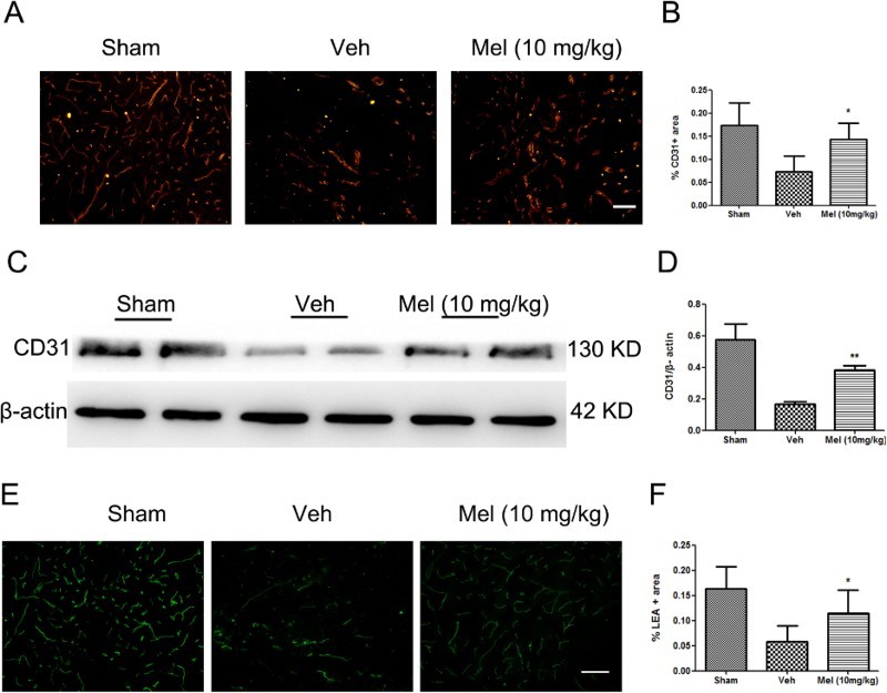
Figure 1.

The effect of melatonin on blood vessels following spinal cord injury (SCI). (A) Blood vessels were visualized by anti-CD31 immunofluorescence in the ventral horn of the spinal cord and were presented for each of the different treatment groups (n = 4). (B) CD31-stained area was quantified in all groups. (C-D) The expression of CD31 was detected by western blot (n = 4), and the relative intensity of CD31 was analyzed in the different treatment groups. (E) Perfused blood vessels were labeled by LEA in the ventral horn of the spinal cord and were presented for each of the different treatment groups (n = 3). (F) LEA-labeled area was quantified in all groups. *P < 0.05 in Veh versus Mel (10 mg/kg), **P < 0.01 in Veh versus Mel (10 mg/kg). Scale bar = 50 μm (Colour online)
(sham group: Sham; vehicle group: Veh; melatonin group: Mel (10 mg/kg))
