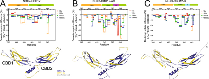
Figure 5.

Effect of Ca2+ binding on NCX2-CBD12 and NCX3-CBD12 dynamics. Deuterium uptake differences between the Ca2+-bound form and the apo form as a function of residue at different time points as indicated are plotted for NCX2-CBD12 (A), NCX3-CBD12-AC (B), and NCX3-CBD12-B (C). The data are also qualitatively depicted on the models of NCX2-CBD12 (A), NCX3-CBD12-AC (B), and NCX3-CBD12-B (C). Regions with lower deuterium uptake in the Ca2+-bound form are in blue, regions with similar deuterium uptake in the Ca2+-bound form and apo form are in yellow, and regions not covered by the detected peptic peptides are in gray.
