
Figure 2.
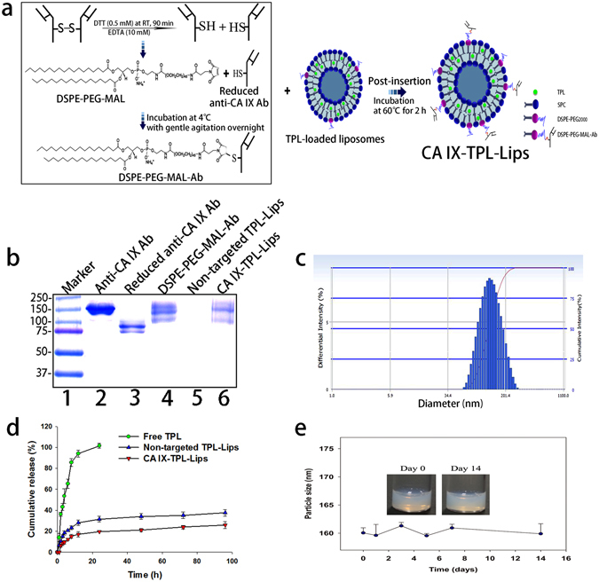
The preparation and characterization of CA IX-TPL-Lips. (a) Illustration of the preparation of CA IX-TPL-Lips; (b) Reducing SDS-PAGE electrophoresis of lane 1: molecular weight size marker, lane 2: Anti-CA IX antibody (Ab), lane 3: Reduced anti-CA IX antibody (Ab), lane 4: DSPE-PEG-MAL-Ab, lane 5: Non-targeted TPL-Lips and lane 6: CA IX-TPL-Lips; SDS-PAGE gel was stained with Coomassie Brilliant Blue R250 to visualize the Ab; (c) Representative particle size distribution of CA IX-TPL-Lips; (d) In vitro release profile of TPL formulations in PBS (pH 7.4); (e) Stability of CA IX-TPL-Lips at 4 °C evaluated by measuring the change in particle size.