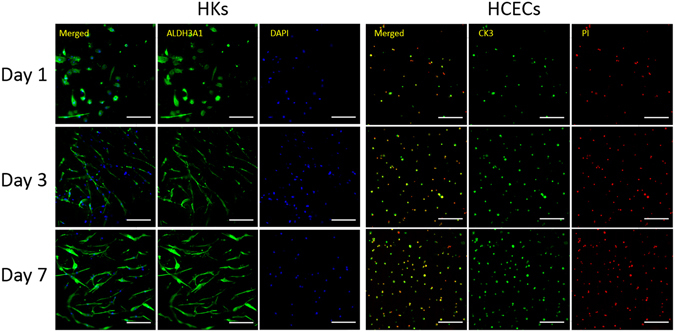
Figure 9.

Protein expression of specific markers in HKs within hybrid scaffolds. Images (left) show the fluorescent staining of ALDH3A1 (green) and nuclei (blue) in HKs after being cultured for 1, 3, and 7 days. Protein expression of specific markers in HCECs seeded on the surface of hybrid scaffolds. Images (right) show fluorescent staining of CK3 (green) and nuclei (red) in HCECs after being cultured for 1, 3, and 7 days. (scale bar, 200 μm).
