
Figure 3.
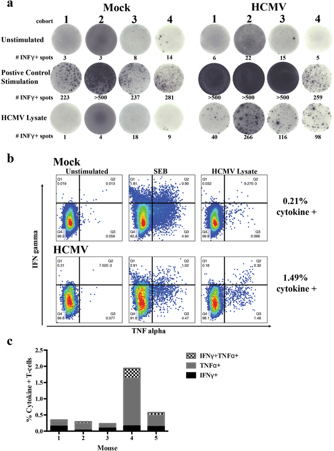
huBLT mice constructed from multiple donor tissues generate HCMV-specific multi-functional T-cell responses. (a) huBLT mice were generated and infected with HCMV as described in Fig. 1. Mice were euthanized at 8 weeks post-infection and splenocytes harvested. Total splenic mononuclear cells left unstimulated or were stimulated with SEB toxin or HCMV viral lysate and cultured on human IFNγ ELISpot plates (MabTech) prior to detection. Mice shown are from four independent experiments using separate donor tissues from Mock-infected (Mock, panel 1) and HCMV-infected (HCMV, panel 2) huBLT mice for each donor. (b) Mock-infected (top) or HCMV-infected (bottom) huBLT mice (cohort 3) were harvested at 6 weeks post-infection and splenocytes harvested. Total splenic mononuclear were unstimulated (panel 1) or stimulated with SEB toxin (panel 2) or HCMV viral lysate (panel 3), incubated with Brefeldin A and stained with human-specific antibodies for flow cytometry analysis of T-cell populations and cytokine production. Samples were gated on viable, human CD45+, CD3+ and CD69+ lymphocytes. (c) Quantification of the cytokine (IFNγ+, TNFα+ or IFNγ + TNFα+) responsive T-cells from five independent huBLT mice analyzed as in (b).