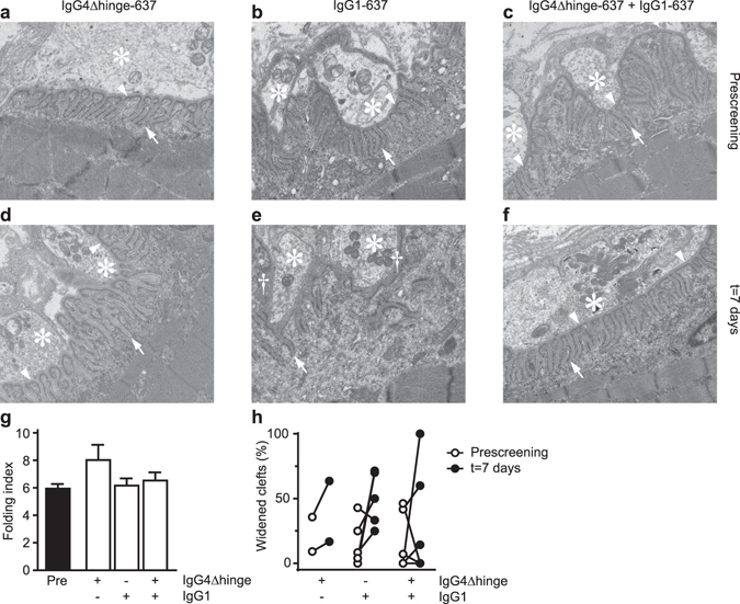
Figure 3.

Electron microscopic analyses of intercostal neuromuscular junctions in rhesus monkeys. Transmission electron micrographs showing representative nerve boutons of 3 animals, either before (‘Prescreening’, a,b,c) or seven days after challenge with IgG4Δhinge-637 (d), IgG1-637 (e) or the combination of IgG4Δhinge-637 and IgG1-637 (f). Each micrograph has the dimension of 5 × 6 µm. Asterisks indicate nerve terminals/boutons and arrowheads point at the (intact) primary synaptic clefts. Arrows point to normal secondary postsynaptic clefts/folds; the daggers in panel e indicate widening of the primary synaptic cleft, where the presynaptic and the postsynaptic membrane were separated from each other. (g) The folding index (length of postsynaptic membrane/length of the corresponding presynaptic membrane), a measure of the degree of postsynaptic folding. (h) Blinded scoring of the normal versus widened synaptic clefts.
