Figure 4.

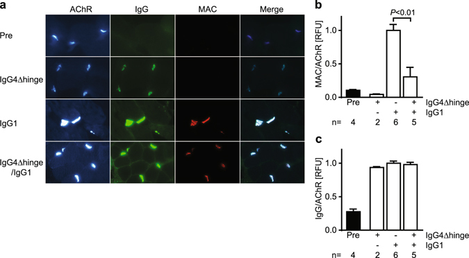
Quantitative Immunofluorescent analysis of NMJ endplates in rhesus monkey intercostal biopsies. Biopsies were obtained from each animal before (“Pre”) or 7 days after antibody challenge with IgG4Δhinge-637, IgG1-637 or the combination of both. (a) Representative photomicrographs from the four groups showing staining of the AChR (detected by alpha-bungarotoxin fluorescence), IgG (IgG + IgG4Δhinge), the membrane attack complex (MAC) and a merged image to show colocalization. Relative fluorescence intensities (RFU) of (b) MAC staining and (c) human IgG staining was normalized with AChR expression in individual endplates. Intensities were quantitated in a total of 589 endplates (5-158 endplates per biopsy) and averaged per biopsy. The number n indicates the number of animals/biopsies analyzed for each condition. Data represent means ± SEM.
