Figure 1.

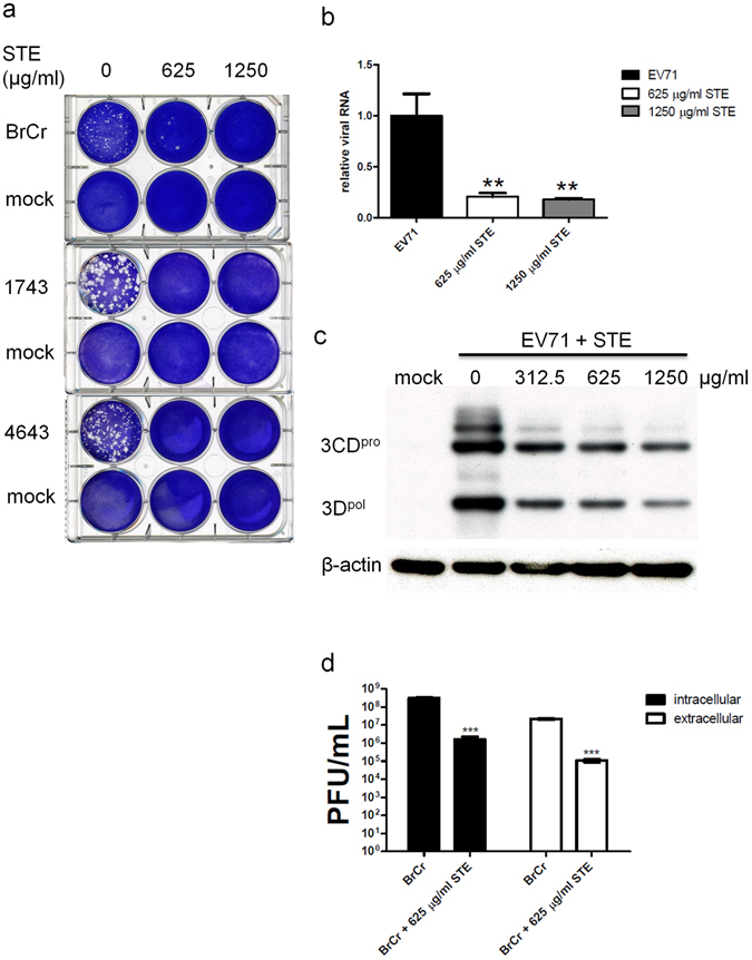
Anti-EV71 activity of STE. (a) RD cells were mock- or infected with 100 PFU of EV71 strains, namely BrCr, 1743 and 4643 for 1 h, and were overlaid with 0.3% agarose in DMEM/2% FBS, which was supplemented with 0, 625, or 1250 µg/ml of STE. After 96 h, the plates were fixed with 10% formalin, and stained with 1% crystal violet solution. Representative cell plates are shown here. (b) RD cells were infected with BrCr at m. o. i. of 0.05 in the absence or presence of 625 and 1250 µg/ml STE for 16 h. The level of EV71 genomic copy was determined by quantitative reverse transcription PCR, and normalized to the level of β-actin. Data are expressed relative to that of untreated cells. The results are means ± SD of three separate experiments. **P < 0.01, vs. infected cells without treatment. (c) RD cells were infected with BrCr at m. o. i. of 5 in the presence of indicated concentrations of STE. Cellular protein was harvested at 6 h p. i., and was subject to western blotting with antibodies to 3D and β-actin. The cropped images of the blots are shown. A representative experiment out of three is shown. (d) RD cells were infected with BrCr as described in (c), and treated without or with 625 µg/ml STE. Extracellular and intracellular viral particles were collected at 9 h p. i. for titer determination. The results are means ± SD of three separate experiments. ***P < 0.0001, vs. infected cells without treatment.
