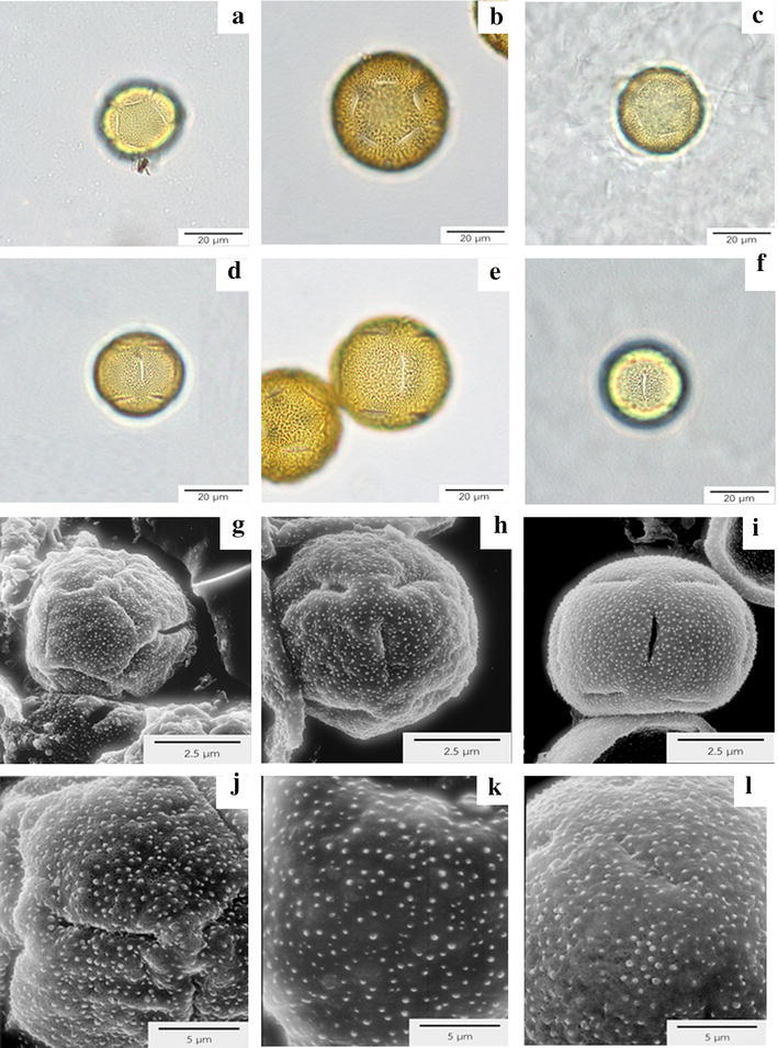
Fig. 3.

Pollen under light microscopy (LM) and scanning electron microscopy (SEM). a–c Polar view under LM of E. alsinoides (a), E. glomeratus (b) and E. nummularius (c), which demonstrates the monad pollen unit and 5-pantocolpate apertures. d–f Monad pollen under LM with 5-pantocolpate apertures at equatorial view of E. alsinoides (d), E. glomeratus (e) and E. nummularius (f). g–i SEM of E. alsinoides (g) in polar view, showing 5-pantocolpate apertures. Polar view under SEM of E. glomeratus (h), equatorial view of E. nummularius (i) presenting 5-pantocolpate apertures. j–l Microechinate ornamentation of E. alsinoides (j), E. glomeratus (k) and E. nummularius (l)
