
Figure 3.
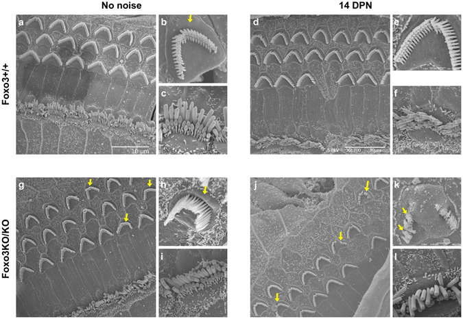
Morphology of inner and outer hair cell stereocilia. Stereociliary bundle morphology of outer (b,e,h,k) and inner hair cells (c,f,i,l) from control Foxo3+/+ mice (a–c) and Foxo3+/+ mice at 14 DPN (d–f) are compared to control Foxo3KO/KO littermates (g–i) and Foxo3KO/KO littermates at 14 DPN (j–l). SEM images are taken from the region corresponding to 12–16 kHz, i.e. within the noise band. In (b) and (h), arrows indicate the apical cuticular region of outer hair cells. In (g) and (j), arrows indicate outer hair cells with stereociliary dysmorphia. In (k), arrows highlight fused stereocilia on a damaged, surviving outer hair cell.