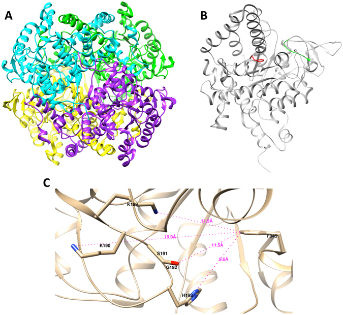
Figure 1.

Structure of MtbICL. (A) Ribbon structure of the homotetrameric MtbICL, with each subunit shown in different color (PDB ID: 1F8I). (B) Chain A of MtbICL tetramer showing the position of Phe345 in red and active site signature sequence (189KKCGH193) in green. (C) Active site cavity of the chain A of MtbICL. The active site residues and Phe345 are marked and labeled. The distances are shown with dotted lines. The structures were visualized with UCSF Chimera.
