Figure 1.

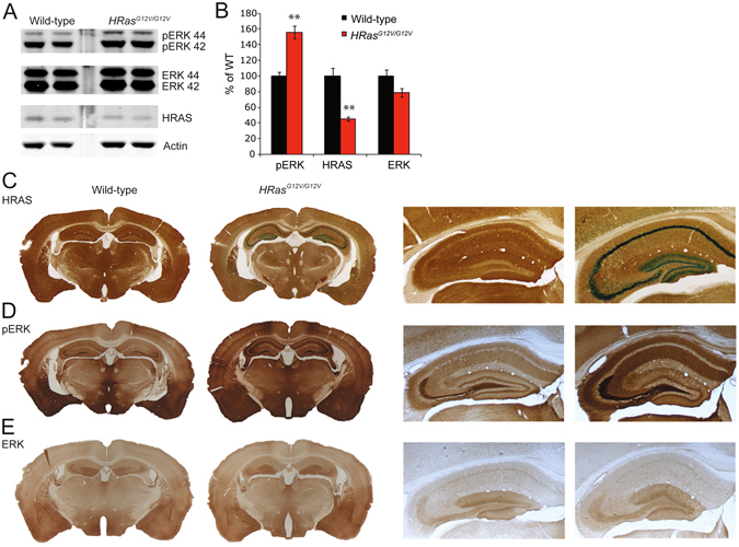
Hyperactivity of RAS-ERK signaling in HRas G12V/G12V mice. (A) Western blots of hippocampal lysates taken from wild-type and HRas G12V/G12V mice using antibodies for HRAS, pERK 42/44 (pERK), ERK 42/44 (ERK) and actin. (B) Quantification of Western blots showing a significant increase in pERK (t 8 = −5.92, P < 0.01) and a significant decrease in expression of HRAS protein in HRas G12V/G12V mice compared to their wild-type littermates (t 8 = 5.34, P < 0.01). Data are presented as mean ± SEM of percentage of WT level set as 100% from 5 mice per genotype (n = 5/genotype). Statistical test: unpaired two-tailed t-test, *P < 0.05, **P < 0.01. (C) Visualisation of β-galactosidase activity of the IRES–β-geo cassette integrated into the mutated HRAS gene (LacZ; blue) combined with an HRAS staining (DAB; brown) indicating successful recombination and expression of HRASG12V, however lower amounts of HRAS protein are seen in HRas G12V/G12V mice compared to WT littermates. Right panel: Zoomed-in image of hippocampus. (D,E) DAB staining using pERK 42/44 and ERK 42/44 antibodies revealed increased levels of ERK phosphorylation in HRas G12V/G12V mouse brain slices compared to WT (D), but no difference in the level of expression and distribution of total ERK between genotypes (E).
