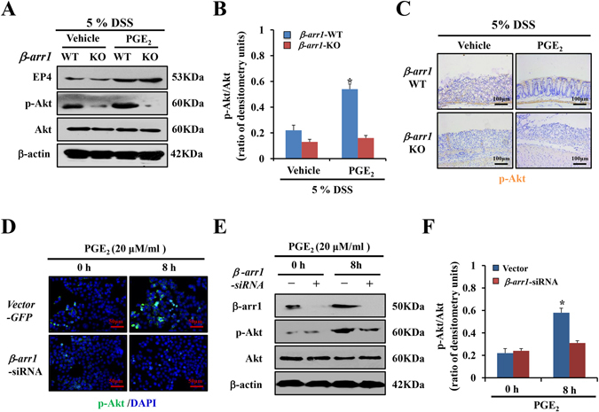
Figure 6.

PGE2/EP4 upregulates β-arr1 mediated Akt signaling in vivo and in vitro. (A) Western blotting of EP4, p-Akt and Akt in colonic mucosa of β-arr1 WT and KO mice treated with DSS with or without PGE2 at the indicated time points. All western blot images are representative images from three independent experiments, and β-actin was used as a loading control. (B) Quantitative analysis of p-Akt/Akt ratio as measured by densitometry scanning of Western blots. Values are expressed as the mean ± SD of three separate experiments, *P < 0.05 versus vehicle. (C) Immunohistochemical staining for p-Akt staining in colonic sections of β-arr1 WT and KO littermates under DSS with or without PGE2 treatment at the indicated time points (brown, ×200, n = 4 per group). (D) Expression of p-Akt in HCT116 cells transfected with control or β-arr1 siRNA after PGE2 treatment. Nuclei were stained with DAPI in blue. Localization of p-Akt was visualized in green. (E) HCT116 cells were stimulated with or without PGE2 (20 μm/ml) for 8 h in the presence of control siRNA or β-arr1 specific siRNA. The lysates were subjected to Western blotting analysis for β-arr1, p-Akt and Akt. All western blot images are representative images from three independent experiments, and β-actin was used as a loading control. (F) Quantitative analysis of the p-Akt/Akt ratio as measured by densitometry scanning of Western blotting. Values are expressed as the mean ± SD of three experiments, *P < 0.05 versus control siRNA group.
