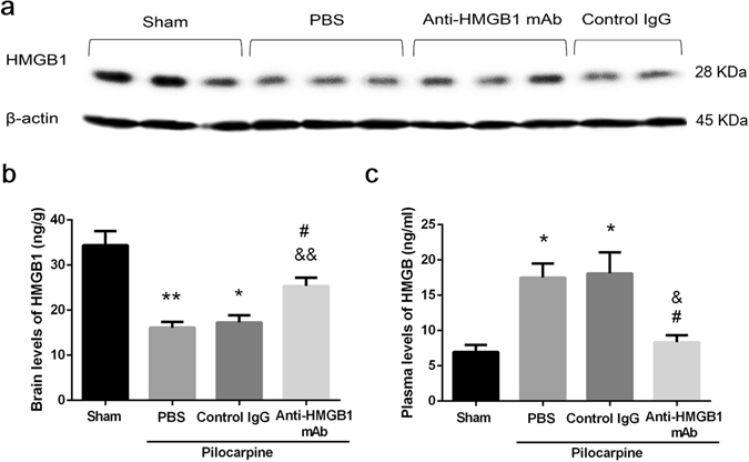
Figure 2.

Dynamic changes of HMGB1 content in brain and plasma in pilocarpine-induced acute status epilepticus. The content of HMGB1 in the cerebrum was measured by Western blotting 4 h after anti-HMGB1, control IgG or PBS injection. The β-actin was used as a reference protein. Representative results are shown for each group of 2–3 mice (a). The brain levels of HMGB1 are shown as the means ± SEM of 9 mice. *p < 0.05, **P < 0.01 compared with the sham control, &&p < 0.01 compared with the PBS control, #p < 0.05 compared with the control IgG group (b). Plasma levels of HMGB1 were determined by ELISA at 4 h after onset of seizure . The results are shown as the means ± SEM of 9 mice. *p < 0.05 compared with the sham control, &p < 0.05 compared with the PBS control, #p < 0.05 compared with the control IgG group (c).
