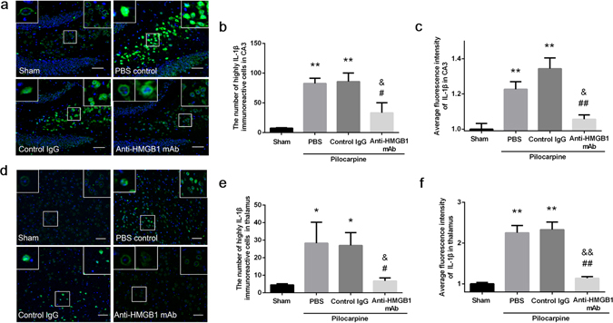
Figure 6.

Expression of IL-1β in the CA3 hippocampal region and the thalamus of mice under acute status epilepticus. Anti-HMGB1 mAb, control IgG or PBS was administered intravenously when mice reach Racine stage 5 by pilocarpine administration and sacrificed 4 h later. The brains were fixed by formalin and paraffin-embedded sections were stained with anti-IL-1β antibody. The hippocampal CA3 (a) and thalamus region (d) were under observation. The number of highly IL-1β immunoreactive cells (b) and the fluorescence intensity (c) were analyzed in hippocampus CA3. **p < 0.01 compared with the sham group. &p < 0.05 compared with the PBS group. ##p < 0.01, #p < 0.05 compared with the control IgG group. The results are shown as the means ± SEM of 8 mice. The number of highly IL-1β immunoreactive cells (e) and the fluorescence intensity (f) were analyzed in thalamus. The results are shown as the means ± SEM of 8 mice. **p < 0.01, *p < 0.05 compared with sham group. &&p < 0.01, &p < 0.05 compared with the PBS group. ##p < 0.01, #p < 0.05 compared with the control IgG group (d). Scale bars equal 50 μm.
