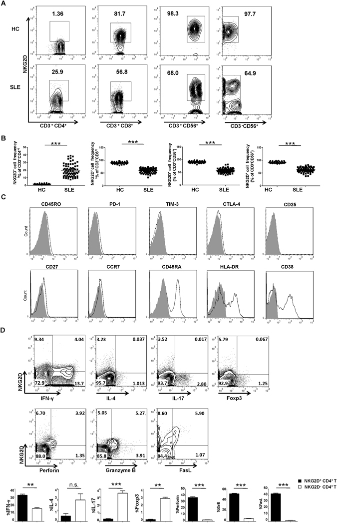
Figure 1.

Identification and phenotypic characterization of NKG2D+CD4+ T cells in SLE. (A) FCM assay of NKG2D expression among CD4+ T, CD8+ T, NK and NKT cells in representative peripheral blood samples from a representative HC and SLE patient. (B) Frequency profiles of NKG2D expression on CD4+ T, CD8+ T, CD3−CD56+ NK and CD3+CD56+ NKT cells in the HCs (n = 46) and SLE patients (n = 66). Each symbol represents one individual. (C) NKG2D+CD4+ T cells were gated from a representative peripheral blood sample of an SLE patient and analyzed for expression of the indicated surface markers (black-lined histograms). Gray-filled histograms represent the staining with control Ab. (D) FCM assay of the expression of the indicated markers in gated CD4 cells from a representative peripheral blood sample of an SLE patient and statistical analysis of the frequency of the of CD4+ T cell subset in PBMCs of SLE patients (n = 30) and HCs (n = 30), respectively. Numbers represent percentages. Each symbol represents one individual; ** P < 0.01, *** P < 0.001. Horizontal lines with bars show the mean ± SD.
