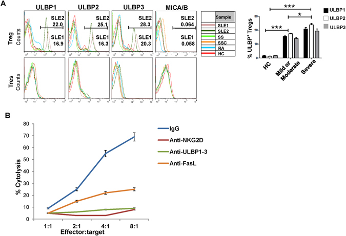
Figure 3.

NKG2D+ CD4+ T cells killed Treg cells had upregulated NKG2DLs in vitro. (A) Induction of ULBP expression on Treg cells by the indicated serum samples. Treg cells were isolated from PBMCs of healthy controls (HCs) and stimulated in vitro by the addition of 10% serum from SLE (n = 10), Sjogren syndrome (SS) (n = 5), systemic scleroderma (SSc) (n = 8), rheumatoid arthritis (RA) (n = 8) patients, or HC (n = 9), respectively, for 18 h, followed by FCM assay. CD4+CD25− T responders (Tres) served as controls. The histograms (left) show original data from one representative experiment. The SLE1 and SLE2 samples are representative serum from patients with mild/moderate (n = 4) and severe (n = 6) SLE, according to SLEDAI index. Bar graphs (right) present the statistical results obtained from three independent experiments with sorted healthy Treg cells stimulated with the indicated serum. * P < 0.05, *** P < 0.001. (B) Freshly isolated Treg cells from HC were stimulated with the serum of SLE patients, labeled with 51Cr and irradiated (25 Gy), co-cultured for 6–8 h with NKG2D+CD4+ T cells from SLE patients, and subjected to cytotoxicity assay, and assessed for the 51Cr-release at the indicated effector:target ratios; the experiment was performed with or without pre-incubation with the indicated neutralizing antibodies. Data represent three independent experiments with Treg cells from HC.
