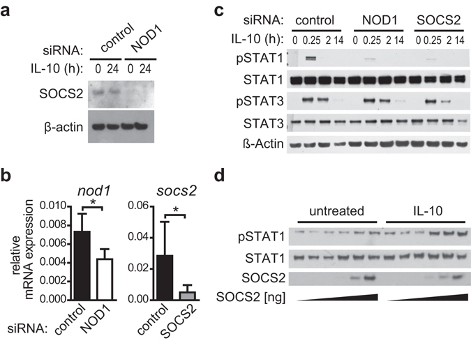
Figure 6.

SOCS2 regulates IL-10-dependent STAT1 phosphorylation. (a) NOD1 silencing and control silencing were performed in iDCs. After 3 days of silencing, iDCs were induced with IL-10 (30 ng/ml) for 24 hours. SOCS2 and β-actin expression was monitored by Western Blot. One of two experiments is shown. (b,c) Immature DCs were transfected with a control siRNA or siRNA directed against NOD1 or SOCS2. (b) 72 hours post transfection, silencing efficiency was analysed by q-RT-PCR. Data represent mean and SD of three independent experiments. For statistical analysis a Student’s t-test was performed. c Transfected cells were stimulated with IL-10 (30 ng/ml) for the indicated time points to analyse STAT1 and STAT3 phosphorylation by Western Blot. To control for equal loading, total STAT protein and β-actin was detected. One representative experiment out of three is shown. (d) HEK293 cells were transiently transfected with expression vectors encoding SOCS2 and the IL-10 receptor subunits IL-10R1 and IL-10R2. 24 hours post transfection, cells were left untreated or stimulated with IL-10 (10 ng/ml) for 20 minutes. STAT1 phosphorylation and SOCS2 expression were analysed by Western Blot. To control for equal loading, total STAT1 was detected. One representative experiment out of three is shown.
