
Figure 7.
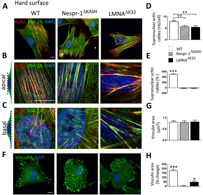
Impaired ability to sustain high external force in Nespr-1ΔKASH and LMNAΔK32 cells. (A) Confocal images of WT, Nespr-1ΔKASH and LMNAΔK32 myoblasts on fibronectin-coated rigid matrix (glass) and stained for F-actin (phalloidin, red) and non-muscle myosin IIA (NM-2A, green). Nuclei are stained with DAPI (blue). Scale bar: 10 µm. (B,C) Zoom-in of actin cytoskeleton in the perinuclear regions. Confocal images were taken at the apical (B) and basal (C) surface of the cell. (D,E) Supranuclear actin cable number on hard surface in WT, Nespr-1ΔKASH and LMNAΔK32 myoblasts. Values expressed as absolute values (D) and as percent changes versus values obtained at 12 kPa (E). Values are means ± SEM. n ≥ 20 in WT, Nespr-1ΔKASH and LMNAΔK32 myoblasts, ***p < 0.01 vs WT. Only significant difference is figured (F–H) Cell matrix adhesions on hard surface. (F) Confocal images of WT, Nespr-1ΔKASH and LMNAΔK32 myoblasts on hard matrix (glass) and stained with antibody against vinculin (green). Nuclei are stained with DAPI (blue). Scale bar: 10 µm. (G,H) Histograms of vinculin area in WT, Nespr-1ΔKASH and LMNAΔK32 myoblasts, expressed as absolute values (G) and as percent changes versus values obtained at 12 kPa (H). At least 50 cells of each type were analysed. *p < 0.05 and ***p < 0.001 compared with corresponding value obtained on 12 kPa substrate. Only significant difference is figured.