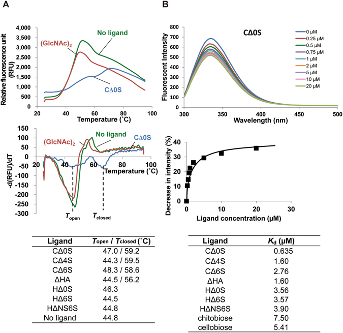
Figure 5.

Affinity of Smon0123 with unsaturated GAG disaccharides. (A) DSF analysis. Upper, fluorescence profile of Smon0123 with C∆0S (green: 0 mM and cyan: 1 mM) or 1 mM N,N′-diacetylchitobiose (red). Middle, negative derivative curve plot derived from the fluorescence profile. Lower, the values of T open and T closed in the presence or absence of various unsaturated GAG disaccharides. (B) Fluorescence spectrum analysis. Upper, wavelength-scanned fluorescence intensity of Smon0123 with C∆0S (blue: 0 μM; red: 0.25 μM; green: 0.50 μM; purple: 0.75 μM; cyan: 1.0 μM; orange: 2.0 μM; lilac: 5.0 μM; pink: 10 μM; and olive: 20 μM). Middle, the relative fluorescence intensity by addition of increasing ligand concentrations was plotted after modification based on volume change in the cuvette. K d was determined using the least-squares method. Lower, dissociation constants of Smon0123 with various unsaturated GAG disaccharides. Chitobiose, N,N′-diacetylchitobiose.
