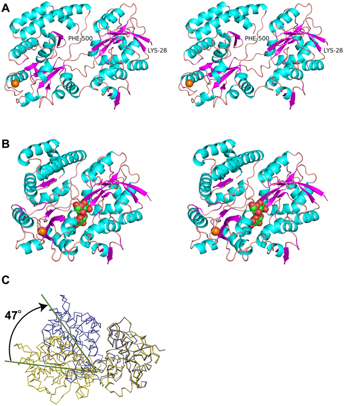
Figure 6.

Overall structures of Smon0123. Structures (stereo-diagram with wall-eyed viewing) of ligand-free Smon0123 (N-28/C-5) (A) and CΔ0S-bound Smon0123 (N-18/C-5) (B). Cyan, α-helices; purple, β-strands; pink, loops and coils. CΔ0S bound to Smon0123 is shown in ball model (green, carbon atom; red, oxygen atom; and blue, nitrogen atom). Orange ball shows the metal ion. Structural comparison with ligand-free Smon0123 (N-28/C-5) (olive) and CΔ0S-bound Smon0123 (N-18/C-5) (blue) (C). Both N-domains of ligand-free and -bound Smon0123 proteins were superimposed. There is a structural difference (47° in angle) in the N- and C-domains between C∆0S-free and -bound Smon0123.
