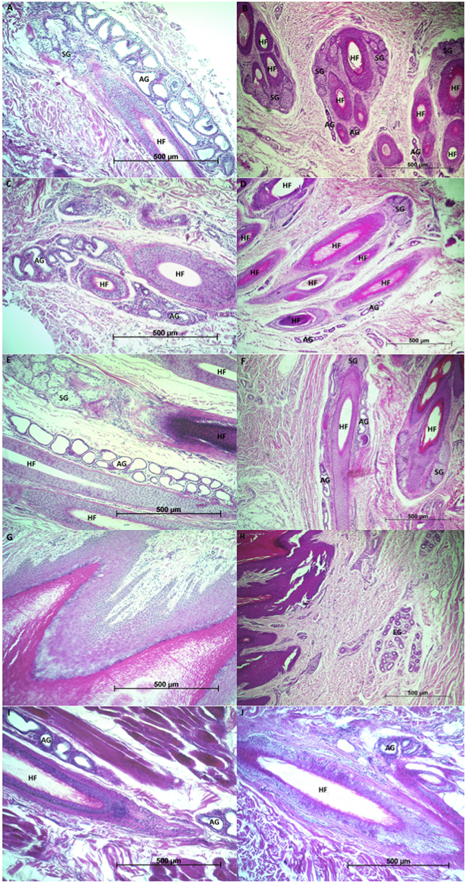
Figure 1.

Histological sections of interdigital skin of front paw in adult male (A), interdigital skin of front paw in yearling male (B), interdigital skin of hind paw in adult male (C), interdigital skin of hind paw in yearling male (D), ventral metatarsal skin in adult male (E), ventral metatarsal skin in yearling male (F), front footpad in adult male (G), front footpad in yearling male (H), and control lip skin (I) and control left shoulder skin (J) in adult male brown bear (Ursus arctos). In interdigital and metatarsal regions (A,B,C,D,E,F) there are hair follicles (HF) visible with apparently more profuse and prominent apocrine sweat glands (AG) in adult male (A,C and E). In sections of control skin of the adult male (I and J), apocrine sweat glands appear less profuse than in sections of the focus areas of the paws. Sections of footpads (G and H) show the stratified squamous keratinized epithelium of the pad with very thick striatum corneum (the darkest shade of stain), and eccrine glands (EG) deeper in the dermis of yearling male (H). Stain: hematoxylin and eosin. Magnification: x100. SG – sebaceous gland.
