Figure 5.

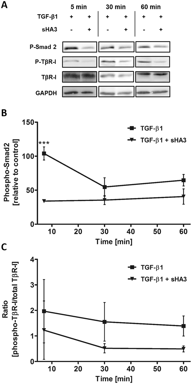
Influence of sHA3 on the TGF-β1-mediated TβR-I and Smad2 phosphorylation. Hs27 fibroblast cells were stimulated with 10 ng/ml TGF-β1 (0.4 nM) alone or pre-formed complexes of TGF-β1 and 100 µg/ml sHA3 (130 µM D.U.) for the indicated time points. Cells were lysed and applied to Western Blot analyses using specific anti-phospho-Smad2, anti-phospho-TβR-I and anti-GAPDH antibodies. Membranes were reblotted afterwards with anti-TβR-I antibody. For every time point a representative blot is shown (A). The time course of TGF-β1-mediated phosphorylation in the presence or absence of sHA3 is plotted for Smad2 phosphorylation (B) and TβR-I phosphorylation relative to unstimulated cells (C). Two-way ANOVA: ***p < 0.001 vs. TGF-β1 only.
