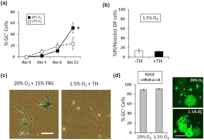
Figure 2.

Hypoxia does neither induce cell death and replicative senescence, nor inhibit OL differentiation. (a) P7 OPCs cultured in PDGF and TH, in either 20% O2 (closed squares) or 1.5% (open squares), were stained for GC after 3, 6 and 12 days, and the % of GC+ cells were counted at each time point. *P < 0.05 (unpaired Student’s t-test, n = 3). (b) P7 OPCs were cultured in PDGF, without TH (-TH, white bar) or with TH (+TH, black bar), in 1.5% O2 for 15 days, and the percentage of dead cells (PI+, Hoechst 33342+ double positive cells) was determined (n = 3). (c) P7 OPCs were cultured in PDGF and TH in 1.5% O2 for 20 days and stained for SA-βGal (left panel); as a positive control, P7 OPCs cultured in 15% FBS in 20% O2 for 20 days to induce replicative senescence were also stained for SA-βGal (right panel). Scale bar: 50 μm. (d) Freshly purified P7 OPCs were cultured in the absence of PDGF for 3 days, in either 20% O2 (upper right panel) or 1.5% O2 (lower right panel), and were labeled with anti-GC antibody (green) and DAPI (blue). Scale bar: 100 μm. Percentages of GC+ cells in 20% O2 culture and 1.5% O2 culture are shown in the graph on the left (n = 3). Note that rat OL differentiation obeying withdrawal of PDGF is not inhibited in 1.5% O2, the extensions of GC+ membrane-like structure around the cell body in 1.5% O2 were comparable with those in 20% O2.
