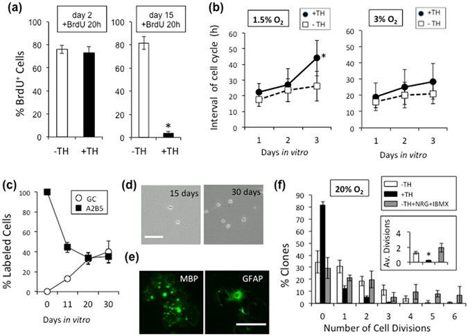
Figure 3.

Hypoxia converts perinatal OPCs to adult-like OPCs in culture. (a) After 2 or 15 days, with TH (+TH; black bars) or without TH (-TH; white bars), the P7 OPCs cultured in 1.5% O2 condition were treated with BrdU for 20 hours and then the % of BrdU+ cells was determined. *P < 0.001. (b) Freshly prepared P7 OPCs were plated in PDGF, with TH (closed circles) or without TH (open squares) and in either 1.5% or 3% O2 cultured for 24 hours, after which they were followed by time-lapse video microscopy. The average time between M-phases was estimated on day 1 (0–23.5 hours), day 2 (24–47.5 hours), and day 3 (48–71.5 hours). At the start of image recording, the following number of cells were analyzed: 42 in 1.5% O2 with TH; 48 in 1.5% O2 without TH; 131 in 3% O2 with TH; 89 in 3% O2 without TH. *P < 0.05. (c) After P7 OPCs were cultured at clonal-density for 11, 20 or 30 days in PDGF, TH, and 1.5% O2, the cells were labeled with anti-GC and A2B5 monoclonal antibodies and the percent of each type of labeled cells was determined. (d) P7 OPCs were cultured in PDGF and TH in 1.5% O2 for 15 or 30 days, and representative fields were examined by phase-contrast microscopy. Scale bar: 50 μm. (e) P7 OPCs cultured for 15 days as in (d) were then either deprived of PDGF for 5 days and labeled for MBP or treated with 10% FBS for 5 days and labeled for GFAP. Scale bar: 100 μm. (f) P7 OPCs cultured in PDGF and TH in 1.5% O2 for 15 days were removed and re-cultured at clonal-density for another 7 days in PDGF in 20% O2 — without TH (white bars), with TH (black bars), or without TH in the presence of NRG1 (50 ng/ml) and IBMX (100 μM) (gray bars). The average numbers of cell divisions are show in the inset (1.24 ± 0.18 without TH; 0.24 ± 0.60 with TH; 1.95 ± 0.60 without TH, but with NRG1 and IMBX). *P < 0.001.
