Figure 6.

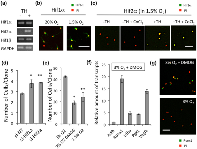
Stabilization of Hifα proteins permits TH-dependent Runx1 gene expression. (a) P7 OPCs cultured in 1.5% O2 in PDGF, with (+) or without TH (−), for 15 days were assayed gene expressions of Hifs by RT-PCR. (b) P7 OPCs were cultured in 20% O2 in PDGF without TH for 7 days, and some were then changed to 1.5% O2 for 20 hours. The cells were labeled with rabbit anti-Hif1α antibodies (green) and PI (red). Scale bar: 50 μm. (c) P7 OPCs were cultured in 1.5% O2 in PDGF with (+TH) or without TH (−TH) for 10 days. The cells were labeled with rabbit anti-Hif2α antibodies (green) and PI (red). To prevent the degradation of Hif2α protein, some cultures were treated with 0.2 mM of CoCl2 65 for last 7 hours. Scale bar: 50 μm. (d) P7 rat OPCs were cultured without TH in 1.5% O2 conditions for 12 days, then co-transfected with anti-Hif1α siRNA or anti-Hif2α siRNA and pMaxGFP. Cells were cultured with TH in 1.5% O2 conditions for another 4 days. The number of GFP+ cells in each clone was counted. si-NT; non-target siRNA, si-Hif1a; anti-Hif1α siRNA, si-Hif2a; anti-Hif2α siRNA. *P < 0.05, **P < 0.01. (e) Freshly prepared OPCs from P7 rat optic nerve were cultured with TH in 1.5% O2 or 3% O2 conditions. For several flasks in 3% O2, DMOG (1 mM) was added. After 7 days, the number of cells in each clone was counted. *P < 0.005, **P < 0.05. (f) P7 rat OPCs cultured in 3% O2 with TH were treated with DMOG (1 mM) for 24 hours. Then qRT-PCR analysis was carried out. Results were presented as the relative amount of transcripts to that of the DMOG free culture using comparative ∆∆Ct method. Actb is an endogenous negative control. Ldha, Pgk1 and Vegfa are HIFs-inducible positive control. The P values of these genes are P < 0.001 (ANOVA with Fisher’s LSD test, n = 3). (g) After 24 hours of DMOG treatment, OPCs (3% O2 + TH) were stained with rabbit anti-Runx1 antibodies (green) and PI (red). Scale bars; 50 μm.
