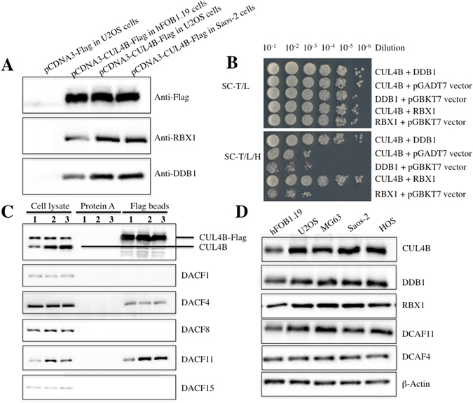
Figure 2.

CUL4B interacted with DDB1, RXB1 and DCAF11 to form a complex in vivo and in vitro. (A) Flag-tagged CUL4B interacted with RBX1 and DDB1 in vivo. The pCDNA3-CUL4B-Flag or pCDNA3-Flag empty vector was transformed into hFOB1.19, U2OS or Saos-2 cells. After 48 h incubation, Flag-tagged proteins were immunoprecipitated with anti-Flag agarose. The pull-down products were analysed by immunoblotting with anti-Flag, anti-RBX1, or anti-DDB1 antibodies. (B) Interactions between CUL4B/DDB1 and CUL4B/RBX1 in yeast. The pGBKT7-CUL4B plasmid was co-transformed with pGADT7-DDB1 or pGADT7-RBX1 into the yeast strain AH109. Growth of the transformed yeast was assayed on media lacking Trp and Leu (top panel) or lacking Trp, Leu, and His (bottom panel). Columns in each panel represent serial 10-fold dilutions. (C) Flag-tagged CUL4B associated with DCAF11 in vivo. The pCDNA3-CUL4B-Flag plasmid was transformed into hFOB1.19 (1), U2OS (2) or Saos-2 (3) cells. After 48 h incubation, Flag-tagged proteins were immunoprecipitated with either protein A agarose or anti-Flag-agarose. The cell lysate and pull-down products were analysed by immunoblotting with anti-CUL4B, anti-DCAF1, anti-DCAF4, anti-DCAF8, anti-DCAF11 or anti-DCAF15 antibodies. (D) DCAF11 was overexpressed in human osteosarcoma cell lines. Cell lysates were applied to WB analyses with specific antibodies against CUL4B, DDB1, RBX1, DCAF11, DCAF4, or β-Actin.
